Ankle Plantar Flexion Soleus Placement
Electrode Placement
Application Instruction by Dr. Lucinda Baker
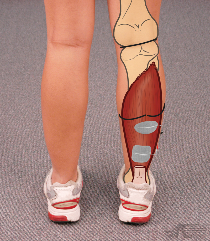
Electrode placement for plantar flexion stimulation soleus activation. The two heads of the gastroc are marked. Using an asymmetric waveform, the negative electrode is placed just below the heads of the gastroc.
The positive electrode is placed above the Achilles tendon. This allows isolated activation of the soleus.
Ankle Plantar Flexion Soleus Placement
Video Instruction
Audio Transcript:
Electrode placement for plantar flexion stimulation soleus activation. The two heads of the gastroc are marked. Using an asymmetric waveform, the negative electrode is placed just below the heads of the gastroc. The positive electrode is placed above the Achilles tendon. This allows isolated activation of the soleus.
During stimulation, the stimulated extremity is slightly un-weighted with the opposite foot placed in front. Heel rise can be seen during stimulation in this position.
Ankle Plantar Flexion Soleus Placement
Muscle Anatomy
Muscles involved in ankle plantar flexion:
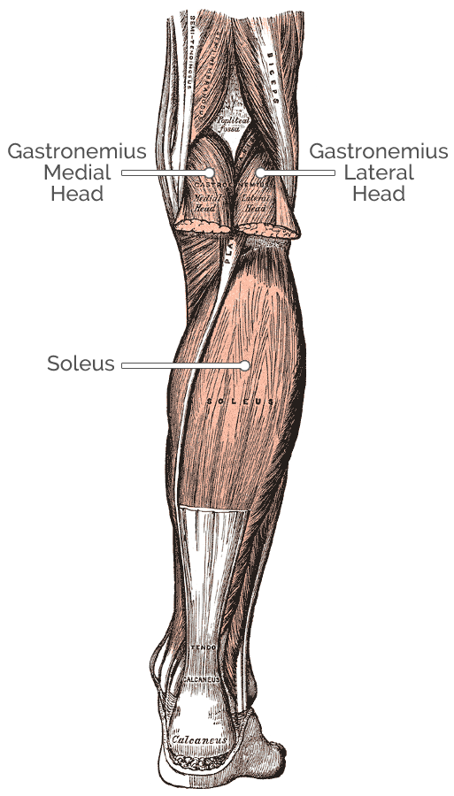
Gastrocnemius
Origin: Superior to articular surfaces of lateral condyle of femur and medial condyle of femur
Insertion: Tendo calcaneus (achilles tendon) into mid-posterior calcaneus
Other actions: Flexes knee
Soleus
Origin: Fibula, medial border of tibia (soleal line)
Insertion: Tendo calcaneus
Plantaris
Origin: Lateral supracondylar ridge of femur above lateral head of gastrocnemius
Insertion: Endo calcaneus (medial side, deep to gastrocnemius tendon)
Other actions: Flexes knee
Flexor Hallucis Longus
Origin: Fibula, posterior aspect of middle 1/3
Insertion: Plantar surface; base of distal phalanx of hallux
Flexor Digitorum Longus
Origin: Posterior surface of the body of the tibia
Insertion: Plantar surface; base of the distal phalanges of the four lesser toes
Tibialis Posterior
Origin: Tibia and fibula
Insertion: Navicular and medial cuneiform bone
Other actions: Foot inversion
Peroneous Longus
Origin: Proximal part of lateral surface of shaft of fibula
Insertion: First metatarsal, medial cuneiform
Other actions: Foot eversion
Ankle Plantar Flexion Soleus Placement
Nerve Anatomy
Nerves involved in ankle plantar flexion:
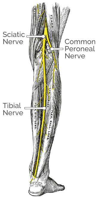
Gastrocnemius
Nerve innervation: Tibial nerve from the sciatic nerve
Nerve root: S1–S2
Soleus
Nerve innervation: Tibial nerve
Nerve root: L5-S1
Plantaris
Nerve innervation: Tibial nerve
Nerve root: anterior rami of S1-S2
Flexor Hallucis Longus
Nerve innervation: Tibial nerve
Nerve root: primarily S1 & S2 secondary L5 nerve roots
Flexor Digitorum Longus
Nerve innervation: Tibial nerve
Nerve root: L5-S1
Tibialis Posterior
Nerve innervation: Tibial nerve
Nerve root: L5-S1
Peroneous Longus
Nerve innervation: Superficial peroneal nerve
Nerve root: L5-S2
Peroneous Brevis
Nerve innervation: Superficial peroneal nerve
Nerve root: L4-S2
